Vai al contenuto

File:Virus Replication large.svg

Contenuti della pagina non supportati in altre lingue.
Wikibooks, manuali e libri di testo liberi.

File originale (file in formato SVG, dimensioni nominali 925 × 852 pixel, dimensione del file: 310 KB)

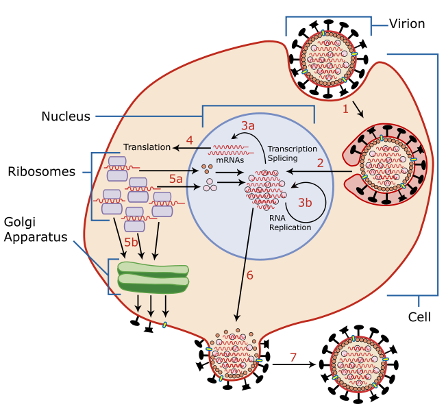
Descrizione A diagram of influenza viral cell invasion and replication.
Data
Fonte Scaled up from Image:Virus Replication.svg by User:YK Times, who redrew from w:Image:Virusreplication.png using Adobe Illustrator. Based on Scheme of Influenza A virus replication, NCBI, artwork by David Hobson, August 6, 2002
Autore User:YK Times
Licenza
(Riusare questo file)
Io, detentore del copyright su quest'opera, dichiaro di pubblicarla con le seguenti licenze:
GNU head È permesso copiare, distribuire e/o modificare questo documento in base ai termini della GNU Free Documentation License, Versione 1.2 o successive pubblicata dalla Free Software Foundation; senza alcuna sezione non modificabile, senza testo di copertina e senza testo di quarta di copertina. Una copia della licenza è inclusa nella sezione intitolata Testo della GNU Free Documentation License.
w:it:Creative Commons
attribuzione condividi allo stesso modo
Questo file è disponibile in base alla licenza Creative Commons Attribuzione-Condividi allo stesso modo 3.0 Unported
Tu sei libero:
  • di condividere – di copiare, distribuire e trasmettere quest'opera
  • di modificare – di adattare l'opera
Alle seguenti condizioni:
  • attribuzione – Devi fornire i crediti appropriati, un collegamento alla licenza e indicare se sono state apportate modifiche. Puoi farlo in qualsiasi modo ragionevole, ma non in alcun modo che suggerisca che il licenziante approvi te o il tuo uso.
  • condividi allo stesso modo – Se remixi, trasformi o sviluppi il materiale, devi distribuire i tuoi contributi in base alla stessa licenza o compatibile all'originale.
Questo tag di copyright è stato aggiunto nell'ambito dell'aggiornamento della licenza dei progetti Wikimedia.
w:it:Creative Commons
attribuzione condividi allo stesso modo
Questo file è disponibile in base alle licenze Creative Commons Attribuzione-Condividi allo stesso modo 2.5 Generico, 2.0 Generico e 1.0 Generico
Tu sei libero:
  • di condividere – di copiare, distribuire e trasmettere quest'opera
  • di modificare – di adattare l'opera
Alle seguenti condizioni:
  • attribuzione – Devi fornire i crediti appropriati, un collegamento alla licenza e indicare se sono state apportate modifiche. Puoi farlo in qualsiasi modo ragionevole, ma non in alcun modo che suggerisca che il licenziante approvi te o il tuo uso.
  • condividi allo stesso modo – Se remixi, trasformi o sviluppi il materiale, devi distribuire i tuoi contributi in base alla stessa licenza o compatibile all'originale.
Puoi scegliere la licenza che preferisci.
Altre versioni

Dettagli

From Scheme of Influenza A virus replication (NCBI):

"A virion attaches to the host cell membrane using Hemagglutinin(HA) and enters the cytoplasm by receptor-mediated endocytosis (STEP 1), thereby forming an endosome. A cellular trypsin-like enzyme cleaves HA into products HA1 and HA2 (not shown). HA2 promotes fusion of the virus envelope and the endosome membranes. A minor virus envelope protein M2 acts as a ion channel making the inside of the virion more acidic. As a result, the major envelope protein M1 dissociates from the nucleocapsid and vRNAs are translocated into the nucleus (STEP 2) via interaction between NP and cellular transport machinery. In the nucleus, the viral polymerase complexes transcribe (STEP 3a) and replicate (STEP 3b) the vRNAs. Newly synthesized mRNAs migrate to cytoplasm (STEP 4) where they are translated. Posttranslational processing of HA, Neuraminidase(NA), and M2 includes transportation via Golgi apparatus to the cell membrane (STEP 5b). NP, M1, NS1 (nonstructural regulatory protein - not shown) and NEP (nuclear export protein, a minor virion component - not shown) move to the nucleus (STEP 5a) and bind freshly synthesized copies of vRNAs. The newly formed nucleocapsids migrate into the cytoplasm in a NEP-dependent process and eventually interact via protein M1 with a region of the cell membrane where HA, NA and M2 have been inserted (STEP 6). Then the newly synthesized virions bud from infected cell (STEP 7). NA destroys the sialic acid moiety of cellular receptors, thereby releasing the progeny virions.

Annotazioni
InfoField
Questa immagine è annotata: Vedi le annotazioni su Commons

Didascalie

Aggiungi una brevissima spiegazione di ciò che questo file rappresenta

Elementi ritratti in questo file

raffigura

Cronologia del file

Fare clic su un gruppo data/ora per vedere il file come si presentava nel momento indicato.

Data/OraMiniaturaDimensioniUtenteCommento
attuale18:26, 16 mar 2008Miniatura della versione delle 18:26, 16 mar 2008925 × 852 (310 KB)Photohound{{Information |Description=A diagram of influenza viral cell invasion and replication. |Source=Scaled up from Image:Virus Replication.svg by User:YK Times, who redrew from w:Image:Virusreplication.png using Adobe Illustrator. |Date=March 5, 2

La seguente pagina usa questo file:

Utilizzo globale del file

Anche i seguenti wiki usano questo file: